hear.com – India’s leading destination for advanced hearing aids & expert care
Cholesteatoma
Symptoms, diagnosis & therapy
The cholesteatoma is a chronic, purulent inflammation of the middle ear caused by the growth of squamous epithelium from the external auditory canal into the middle ear. Usually the eardrum separates the middle ear and the outer ear canal. In healthy people, the middle ear is lined with mucosal epithelium and the external auditory canal with squamous epithelium.
If this squamous epithelium gets into the middle ear for various reasons (e.g. hole or tear in the eardrum), this can lead to constant inflammation. This in turn can cause permanent destruction of the bone in the area of the tympanic cavity (middle ear) and the temporal bone. This is why one speaks of “chronic bone dilatation” (otitis media cholesteatomatosa).
Table of Contents
- Forms of cholesteatoma
- Cholesteatoma symptoms
- Cholesteatoma Diagnosis
- Cholesteatoma OP
Three forms of cholesteatoma
1. Congenital cholesteatoma
This form of cholesteatoma is very rare and arises from the fact that squamous cells remain in the middle ear during the development of the embryo. The eardrum is intact here.
2. Primarily acquired cholesteatoma or retraction cholesteatoma
The reason for this form of cholesteatoma is a ventilation disorder in the middle ear, often caused by insufficient tube permeability. The tube is a connecting tube between the middle ear and the nasopharynx. This ventilation disturbance leads to a negative pressure in the middle ear and through this negative pressure the eardrum in turn forms a so-called retraction pocket (retraction of the eardrum). This usually happens in the area of the pars flaccida of the eardrum or also called Shrapnell’s membrane. The reason for this is that, unlike other areas of the eardrum, the pars flaccida is very flaccid. Squamous epithelium collects in this retraction pocket and after a few years it ultimately becomes a cholesteatoma.
3. Secondary acquired cholesteatoma
The reason for this type of cholesteatoma is an existing marginal eardrum defect over which the epithelium advances into the middle ear.
Cholesteatoma Symptoms
Usually, the cholesteatoma initially presents itself as a chronic otitis media:
- There is a foul-smelling ear flow (fetid otorrhea)
- Increasing hearing loss (hyperacusis)
- In addition, there is pressure in the ears
In more advanced stages, there are also more serious symptoms such as balance disorders (possible symptom: vertigo) when the cholesteatoma encroaches on the inner ear or the organ of equilibrium.
Another symptom of cholesteatoma is facial paralysis when the facial nerve is affected, due to its close proximity to the middle ear.
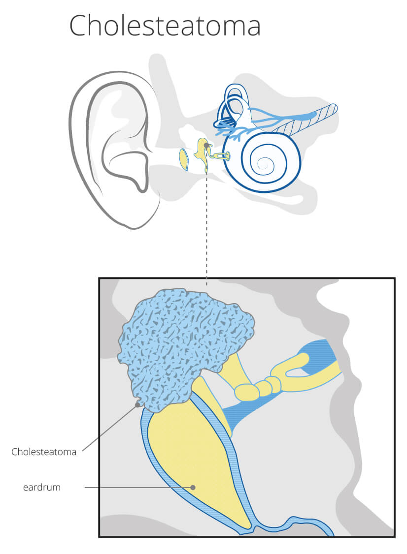
Cholesteatoma
What does a cholesteatoma look like?
Diagnosis
The diagnosis is usually confirmed using three different methods:
1. Ear microscopy
Typical findings here are an eardrum defect, white epithelial scales and possibly bone erosion (progressive destruction) of the wall of the ear canal near the eardrum.
2. CT examination (computed tomography)
Here the exact extent of the bone destruction can usually be determined very well.
3. Hearing test (tone audiogram)
Usually one finds conductive hearing loss in cholesteatoma patients, i.e. a hearing disorder that only affects the outer/middle ear. However, if the cholesteatoma is so advanced that the inner ear is already affected, a so-called sensorineural disorder is present.
Therapy: Cholesteatoma OP
If you want to achieve complete healing of the cholesteatoma, the only option is surgery. Here all inflammatory and destructive foci in the mastoid and tympanic cavity must be radically removed. It is only in the second step, usually during a second operation, that the aim is to improve hearing. This is usually achieved through what is known as a tympanoplasty. The eardrum and, if necessary, the ossicular chain are restored.
If facial paralysis or nystagmus (eye twitching, when the inner ear is affected, more precisely the organ of equilibrium) suddenly occurs, an operation is inevitable.
Conservative therapy, i.e. non-surgical treatment, can only be used in preparation for an absolutely necessary operation. Similar to a simple chronic otitis media, local measures (antibiotic ear drops) or “systemic” antibiotic therapy are used.