hear.com – India’s leading destination for advanced hearing aids & expert care
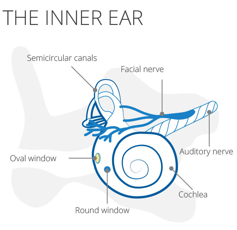
The inner ear
The inner ear is a structure in our skull, more precisely in the temporal bone, i.e. the lateral skull bone. It consists of a membranous and bony labyrinth and is filled with a potassium-rich liquid, the endolymph. Surrounded by the perilymph (sodium-rich liquid), the membranous labyrinth lies within the bony labyrinth. The inner ear is connected to the tympanic cavity, part of the middle ear, through the round and oval windows, two bone openings.
The inner ear is one of our most important sensory organs and houses the anatomical components of the sense of hearing and balance. For these two components of sensory perception, a distinction is made between two separate organs in the inner ear.
Table of Contents
- Our hearing organ
- How does hearing work?
- The hair cells in the inner ear
- The organ of equilibrium
- Inner ear diseases
Inner ear
The hearing organ
The cochlea is responsible for the hearing sensation. This consists of three different courses:
- The Scala vestibuli
- The Scala tympani
- The Cochlear duct
These three courses are separated from each other by membranes. The scala vestibularis merges into the scala tympani at the helicotrema, the tip of the snail. The basilar membrane lies between the scala vestibularis and the scala tympani. The Reissner membrane pulls from the bony wall of the scala vestibuli to the center of the basilar membrane and thus separates the cochlear duct (or the scala media) from the scala vestibuli.
The organ of Corti, in which the actual conversion of the sound stimuli takes place, rests on the basilar membrane within the cochlear duct. This structure, named after the Italian anatomist Alfonso Corti, is the carrier of hair cells (see below), the sensory cells of the inner ear.
The tips of the hair cells (stereovilli) protrude into the tectorial membrane, a gelatinous, inert mass. If the basilar membrane is deflected by vibrations, the stereocilia of the hair cells are bent and an electrical stimulus is generated. A stimulus transduction takes place: a conversion of a mechanical into an electrical (nerve) stimulus.
How does hearing work?
Strictly speaking, a tone is a sound wave. If this sound wave hits our ear, it functions as a funnel and forwards it to the eardrum, which is set in vibration.
These vibrations are then conducted via the ossicles (hammer, anvil and stapes) to the oval window, where they are transmitted to the perilymph in the scala vestibuli.
The sound wave now propagates as a traveling wave in the perilymph to the helicotrema and is then conducted back to the snail base via the scala tympani causing the basilar membrane to be deflected, the stereocilia of the hair cells snap off and trigger an electrical stimulus. This leads to action potentials, which are transmitted to the brain via the auditory nerve, the vestibulocochlear nerve (8th cranial nerve), and processed there.
The hair cells in the inner ear
Specialized receptor cells known as hair cells are found in both the auditory and vestibular organs. However, they have different tasks in the various organs. These are secondary receptors that convert mechanical stimuli into nerve stimuli and thus belong to the class of mechanoreceptors. A hair cell consists of its cell body and the structures that give it its name at its upper end, a cilia (which regresses in the cochlea after birth) and several stereovilli .
The stereocilia are connected to one another by “tip links”. If it is deflected, potassium flows in and the cell is depolarized. As a result, messenger substances are released into the synaptic gap in the lower part of the cell and the following interneuron is excited.
This excitation is then passed on to the brain via the 8th cranial nerve, where it is offset.
The organ of equilibrium
The sense of balance is mediated by the vestibular apparatus. This consists of two atrial sacs, sacculus and utriculus, and three semicircular canals.
Sacculus and utriculus measure the acceleration of our body. The three semicircular canals are located differently in space and can be divided into anterior, posterior and horizontal semicircular canals.
Based on the excitation in the various semicircular canals, the brain can calculate the position of the body. The semicircular canals are filled with endolymph. Your sensory cells, the hair cells, are located inside the cochlea. Their tips protrude into a gelatinous dome, the cupula.
If the head rotates (with or without the rest of the body), the head rotates with the cupula against the lazy endolymph. So the endolymph flows backwards, so to speak.
This deflects the cupula, kinks the hair cells and creates an electrical stimulus . Depending on which of the semicircular canals this stimulus originated in, the brain can calculate the position of the body in space.
Inner ear diseases
Such a complex system is prone to failure. Therefore, there is a variety of diseases at different levels of the structures of the ear:
- Increased sound pressure can damage the hair cells in the inner ear and lead to inner ear hearing loss. In such a case, hearing aids can be used to counteract the hearing loss.
- Inflammation can cause damage to the middle and inner ear and, by destroying important structures, lead to hearing loss or even deafness.
- Benign and malignant tumors can also lead to hearing loss. An example is the acoustic neuroma, which is a benign tumor that originates from the Schwann cells of the 8th cranial nerve and grows in a displacing manner.